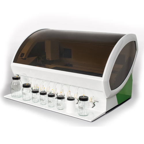
Incubation of a separate membrane strip for each serum sample.
The total time for performing the Western blot test is about 115 minutes.
All incubation steps proceed at room temperature.
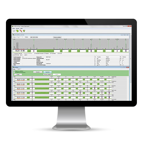
The bands are assigned according to a lot-specific evaluation matrix.
A separate lot for each electrophoresis gel helps avoid errors in the assignment of the bands.
Every test kit contains a membrane strip of the same lot with a positive reference serum. Therefore, there is no need to incubate a positive control serum.
Pre-numbered membrane strips to prevent confusion.
Laborious labeling is not necessary.
Staining of the control band at the bottom of the strip showing correct completion of the individual incubation steps for each membrane strip
Easily and reliably differentiate positive and negative reactions. The intensity of the antigen bands correlates with the antibody titer.
The Western blot is the method of choice when the objective is to confirm or differentiate positive results obtained in a screening test (indirect immunofluorescence or microplate ELISA).
Test principle
The solid phase includes membrane strips containing electrophoretically separated antigen extracts. Therefore, the position of the proteins depends on their respective molecular masses.
If the sample is positive, specific antibodies in the diluted serum sample attach to the antigens coupled to the membrane.
In a second incubation step, the attached antibodies react with AP-labeled anti-human antibodies.
In a third step, the bound antibodies undergo staining with a chromogen/substrate solution which is capable of promoting a color reaction. Thus, an intense dark band at the line of the corresponding antigen appears if the serum sample contains specific antibodies.
Evaluating the band patterns on the incubated membrane strips involves differentiating non-specific from specific antibodies. Moreover, the number and intensity of the specific bands are decisive for the result “positive/negative”.